
Implant Surgery & Support
Bone grafting types & materials
When jawbone has lost volume, grafting restores the foundation an implant needs. The type of graft and the material both depend on where the deficiency is and how large.
Types of graft
Socket Preservation
Performed immediately after an extraction, placing graft material into the empty socket to prevent the rapid resorption that otherwise follows. The most common and most straightforward form of grafting — and the cheapest way to protect a future implant site.
Ridge Augmentation
Where the ridge has already lost width or height, graft material is placed along the deficient area, usually with a membrane, and allowed to heal over three to six months.
Sinus Lift
Where the upper back jaw lacks height beneath the maxillary sinus, the sinus membrane is elevated and graft material placed beneath it. One of the most frequently performed grafting procedures in implant dentistry.
Block Graft
For severe deficiency, a block of bone is harvested from elsewhere in the jaw or sourced from a bone bank. Reserved for significant defects where particulate grafting alone would not be enough.
Graft materials explained
Autograft — Your Own Bone
Harvested from another area of your jaw. The most biologically compatible option, because it contains your own living cells. Used mainly in block grafts and smaller augmentations.
Allograft — Processed Donor Bone
Processed human bone from a tissue bank, acting as a scaffold your body gradually replaces with new bone. Widely used, well documented, and avoids a second surgical site.
Xenograft — Animal-Derived
Processed bovine bone is the most common. It provides a stable scaffold with excellent volume maintenance, which makes it particularly useful in sinus lifts and ridge augmentations.
Synthetic — Laboratory Made
Calcium phosphate ceramics and bioactive glass avoid biological tissue entirely. The option for patients who prefer a tissue-free graft on principle.
How grafting is planned here
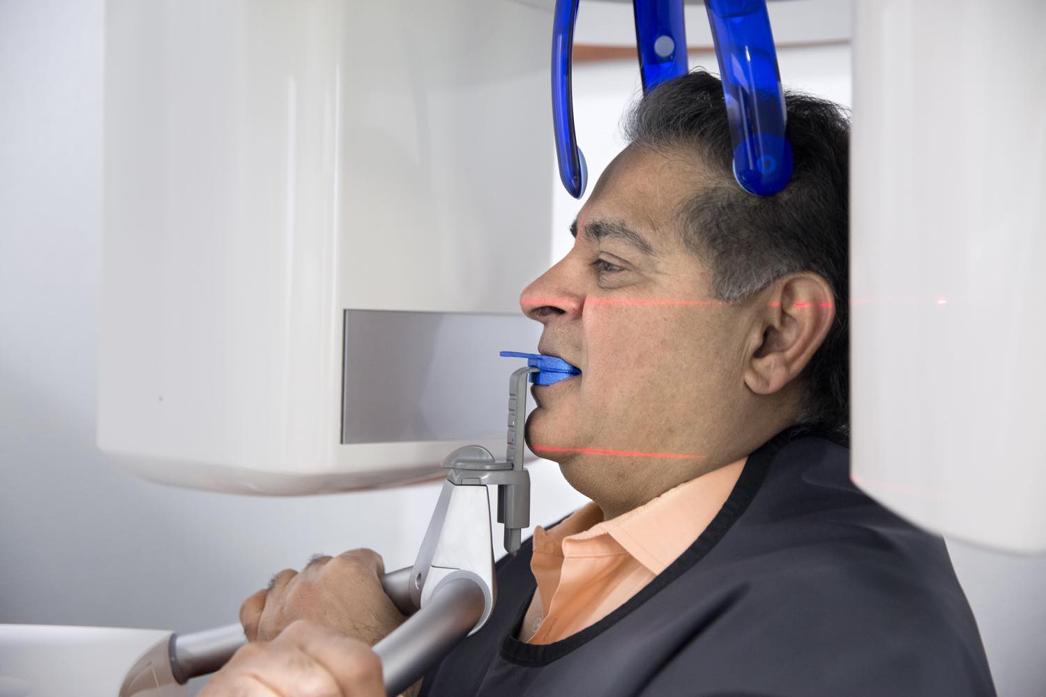
Bone grafting is performed by Dr. Claudio Tocchio, oral and maxillofacial surgeon, and planned in coordination with Dr. Raj Singh based on 3D CBCT imaging showing the exact dimensions of the deficiency.
After grafting, the material integrates with your natural bone over three to six months. Once the graft has matured — confirmed by follow-up imaging rather than assumed — the implant phase proceeds.
Questions about your procedure?
Book a consultation to find out which graft, if any, your case actually requires. Call (905) 479-7777.