Technology & Patient Resources
AI in dentistry: what the software actually does
What these tools are used for in planning, what published reviews say they do reliably, and where the evidence does not yet support relying on them.
Three places it helps
Diagnostics
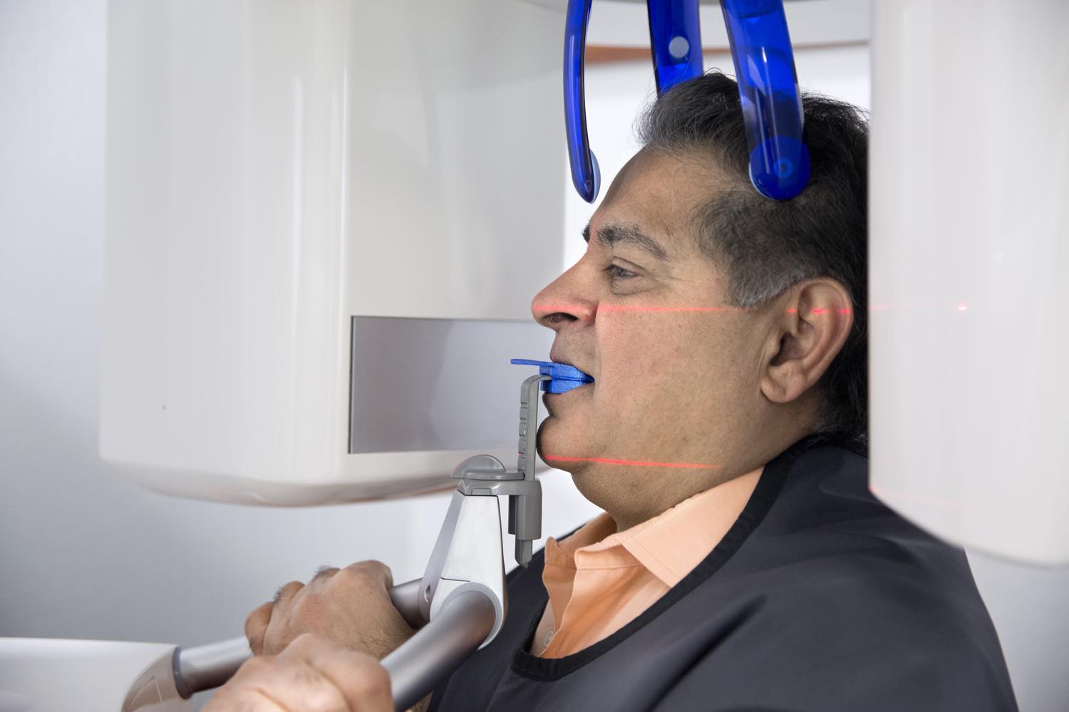
AI algorithms analyse CBCT scans and panoramic radiographs to detect pathology, assess bone density, and identify anatomical structures such as nerve canals and sinus floors — marking areas for a clinician to look at again.
Treatment Planning
In computer-guided surgery, AI tools suggest optimal implant positions based on available bone, the planned restoration, and anatomical constraints. This is particularly valuable in complex cases: full-arch work and situations with limited bone.
Post-Operative Monitoring
Analysis of follow-up radiographs detects early signs of bone loss around implants, identifying patients at risk before symptoms emerge — which is precisely when peri-implantitis is still reversible.
What it does not do
It does not decide. An algorithm can propose an implant position that is geometrically optimal and still be wrong for the patient, because it does not know how hard they grind, what their oral hygiene is realistically like, or what they actually want from treatment.
The technology supports Dr. Raj Singh's clinical expertise rather than replacing it. Forty-plus years of implant experience is the thing that decides whether a suggestion is sensible — and, importantly, when the right answer is not to place an implant at all.
What the reviews say it does reliably
Published reviews report reasonably consistent performance on narrowly defined image tasks. These are the ones worth knowing about, because they are the ones with evidence behind them.
- Finding and numbering teeth, and outlining them on an image. It also spots teeth that are missing, impacted or extra.
- Screening for decay on bitewing and periapical images.
- Measuring bone loss around teeth, which is the measurement that tracks gum disease over time — gum disease.
- Locating dark areas at the tip of a root, which is how an infection shows up on an image.
- Placing orthodontic landmarks and taking the measurements from them — orthodontics.
- Improving an image, sorting a list, and comparing today's image with the last one. Change over time is often the finding.
All of these are measurement and detection tasks with a clear right answer. That is where software is genuinely good.
What the evidence does not support
This is the part usually missing from anything written about AI in dentistry, and it is the more useful half.
- There is little evidence that it improves outcomes. Few studies show it improving treatment decisions or patient outcomes in real practice. Most of the accuracy figures come from old datasets, tested inside the same institution that built the model.
- Performance often drops on images from elsewhere. A model that does well on the machine it was trained on can do much less well on images from another clinic. Different scanner, different settings, different result.
- Many studies use small or hand-picked datasets from a single centre, which flatters the result.
- The reference standard is often one clinician's opinion. Experts do not always agree about what an image shows. So the accuracy figure depends on whose reading was counted as correct.
- High accuracy is not the same as clinically safe. A false positive leads to investigation of something that was not there.
- Performance does not generalise between tasks. Software that outlines teeth well does not therefore diagnose disease.
- It cannot fix a bad image. Overlap, distortion and poor positioning are properties of the radiograph. No amount of processing removes them.
How we actually use it
Descriptively, and without overstating it. These tools are part of the planning software we use for computer-guided implant surgery and inside the imaging software. They outline anatomy on the scan. They mark structures such as nerve canals and sinus floors for us to look at. And they take measurements faster and more evenly than doing it by hand.
A clinician reviews every finding that matters, alongside the examination and the rest of your records. Nothing counts as a diagnosis just because software produced it.
The fair summary in 2026 is this. It is promising for detection, measurement and sorting. The evidence is not strong enough to treat what it produces as settled. We would rather tell you that than market it.
For how the wider digital chain fits together, see digital workflows and our technology.
Sources
The statements above come from published reviews of artificial intelligence in dental imaging. Those reviews describe the evidence as limited in three ways: how well models transfer between clinics, how small the datasets are, and whether outcomes actually improve. Where that is the case, this page says so instead of quoting an accuracy figure without its caveats. Reviewed September 2026.
Common questions
Does AI diagnose my x-rays?+
No. It marks and measures. Every finding that matters is reviewed by a clinician in the context of the examination and the rest of your records. Nothing is treated as a diagnosis because software produced it.
Is it more accurate than a dentist?+
That is not what the evidence shows. Reviews report good performance on narrow detection and measurement tasks, mostly on retrospective data from the institution that built the model, and little prospective evidence that outcomes improve.
Does using AI make my implant more likely to succeed?+
We cannot claim that. Reviews are explicit that few studies demonstrate improved treatment decisions or patient outcomes in real practice. What these tools do is measure and mark faster and more consistently than by hand.
What is it actually good at?+
Finding and numbering teeth, outlining anatomy, screening for decay on bitewing and periapical images, measuring bone loss, locating dark areas at root tips, placing orthodontic landmarks, and comparing today's image with a previous one.
Can software make up for a poor-quality x-ray?+
No. Overlap, distortion and poor positioning are properties of the image itself. Processing cannot remove them, which is why the image is retaken instead.
Have a question?
Book a consultation and see how your case is planned. Call (905) 479-7777.